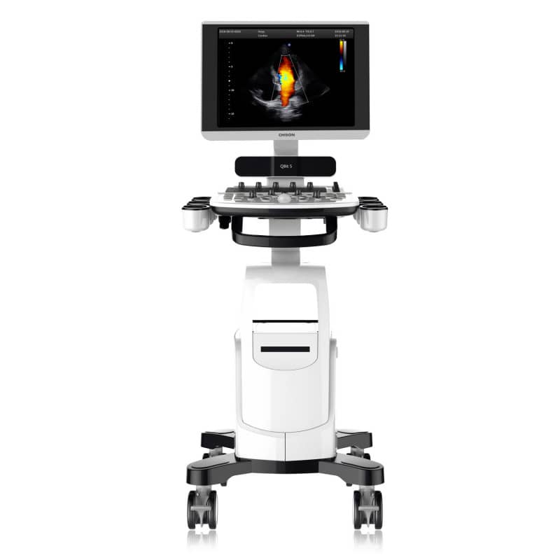
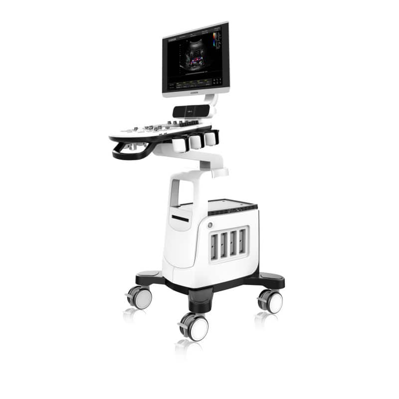
QBit 5 Ultrasound Machine with battery
QBit 5 Ultrasound Machine with battery
Couldn't load pickup availability
- Digital color Doppler ultrasound system
- With high-resolution 19" LED monitor
- FHI, X-cotrast, Q-flow, Q-beam, Q-image
- 32G integrated memory
- Linear & convex probe included
QBit5 Ultrasound Machine
The Chison QBit5 Ultrasound Machine is a digital color Doppler ultrasound system specially designed for demanding medical professionals. The high-resolution 19-inch LED monitor offers precise image display with adjustable brightness and allows detailed diagnosis without loss of image quality thanks to the full-screen mode. The intuitive workflow, simplified alphanumeric keyboard with shortcut keys, and easy-to-use central control ensure efficient operation.
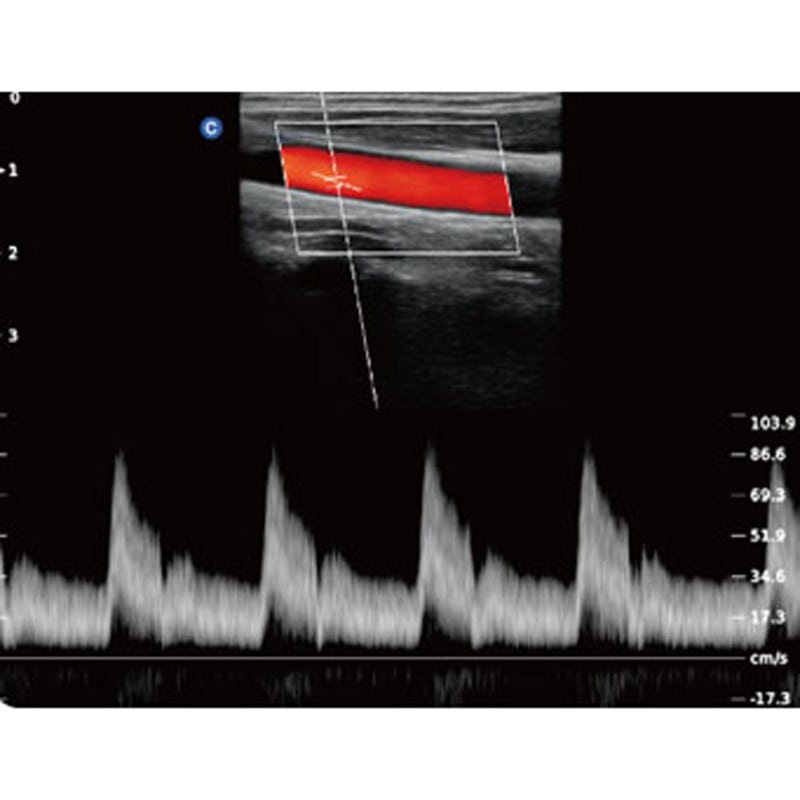
The system employs advanced technologies like FHI, X-contrast, Q-flow, Q-beam, and Q-image to ensure optimal image quality. X-contrast adjusts contrast resolution on three levels based on tissue differences. FHI uses varied transmission and reception methods to maximise resolution for different patient sizes, improving image quality, especially in obese patients. Q-beam delivers superior images and a higher frame rate, enhancing diagnostic reliability and efficiency. Q-flow, an adaptive color detection technology, distinguishes between tissues and adjusts the color signal, improving sensitivity in low-speed flows for more precise diagnostics.
The extensive selection of probes allows a wide range of applications in internal medicine, gynaecology & obstetrics, orthopaedics, paediatrics and urology. The integrated speaker with adjustable volume enhances the system's audio functionality.
Product Details
- QBit5 Ultrasound Machine from CHISON
- Digital color Doppler ultrasound system
- With high-resolution 19" LED monitor (brightness adjustable)
- Equipped with an integrated speaker (volume adjustable)
- Groundbreaking technologies: FHI, X-contrast, Q-flow, Q-beam, Q-image
- Enables an intuitive and intelligent workflow
- Simplified alphanumeric keyboard with shortcut keys
- Easy-to-use central control
- Extensive probe selection allows comprehensive application spectrum
- Full-screen mode without loss of image resolution
- Optimal detail representation for better diagnosis
- Application of a graph facilitates hip orthosis diagnostics
- HIP Graph shows the different degrees of hip deformities (I, II, D, IIIa, IIIb)
- Auto IMT function (optional) facilitates intima measurement
- Optimal needle representation in tissue with Super Needle
- For internal medicine, gynaecology & obstetrics, orthopaedics, paediatrics and urology
- Practical indicator light shows the operational status
- 32G integrated storage
- Includes linear (D7L40L) & convex probe (D3C60L)
- Comes with a smooth-running trolley with 4 lockable wheels
- Available with or without battery
- Manufacturer's warranty: 2 years (battery & probes 6 months)
Indications
Designed for examination in the areas of foetus, abdomen, paediatrics, small organs (breast, thyroid, testes), adult brain, heart (adults, children), musculoskeletal system (conventional, superficial), peripheral vascular diseases, as well as transoesophageal, transrectal, transvaginal and urological examinations.
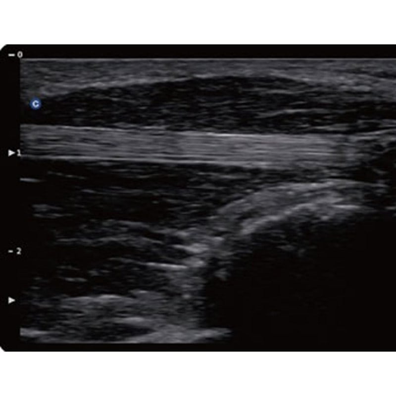
Image Processing Technologies
- SRA (Speckle Reduction Algorithm)
- Compound Imaging (CI)
- Q-Image
- Q-flow
- X-contrast
- Q-beam
- FHI
| X-contrast | - Adjustable contrast resolution on 3 different levels according to tissue differences - Adjustable settings: increase, normal, suppress |
| FHI | - Uses different transmission & reception methods for various patient sizes - More advanced than conventional THI application for better image harmonisation - Helps improve examination results, especially in obese patients |
| Q-beam | - Uses quad-beam method, as opposed to dual-beam, to deliver significantly better images - Higher frame rate ensures better diagnostic reliability and efficiency |
| Q-flow | - Adaptive colour detection technology - Automatically distinguishes between different tissues & adapts the colour signal - Strong improvement in colour sensitivity of low-speed flow |
Imaging Modes
B, 2B, 4B, B/M, M, CFM, B/BC, PW, CW, Colour M, TDI, ECG (optional), PD, Directional PD, Duplex, Triplex, Trapezoidal Image, 2D Steer, Chroma B/M/PW, HIP graph, full-screen, Super Needle (optional), Auto IMT (optional), DICOM, HD 3D (freehand 3D)
Ultrasound Probes
| Convex Probe D3C60L | Linear Probe D7L40L | |
| Application | Abdomen, obstetrics, gynaecology, thorax |
Vascular, small organs, nerves, MSK, paediatrics, orthopaedics, thorax |
| Central Frequency | 3.5 MHz | 7.5 MHz |
| Number of Elements | 128 | 128 |
| Contact Surface | 73 x 16 mm | 44 x 11 mm |
| Operating Modes | B, C, D, M, CPA, DPD | B, C, D, M, CPA, DPD |
| Radius | 60 mm | 40 mm |
| Field of View | 69° | 3.96 cm |
| Scan Depth | 3.2-36.7 cm | 1-14.8 cm |
Connections
| Right Side | 2x probe connectors (standard) 2x USB ports on the control unit |
| Rear | 4x USB ports 1x VIDEO OUT port 1x DVI port 1x VGA port 1x remote control port 1x ECG port 2x footswitch ports 1x LAN port 1x S-Video port |
Share