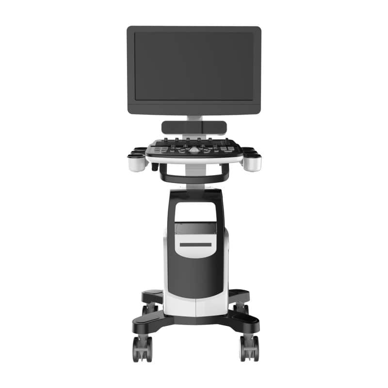
QBit 7 Ultrasound System
QBit 7 Ultrasound System
Couldn't load pickup availability
- Digital color Doppler ultrasound system
- 90° tiltable 21.5” LED screen
- Battery life of up to 80 minutes
- Advanced image processing technologies
- Convex & linear probe included
QBit 7 Ultrasound System
The QBit 7 ultrasound system from CHISON is an advanced colour Doppler ultrasound system specifically designed for medical professionals. It features a 21.5-inch LED display that can swivel 90°, providing flexible viewing angles. The system is equipped with an intuitive user interface with backlit buttons and a 45° swivelling control panel. The built-in speaker and instant print output via the front paper output make it particularly easy to use.
Among the standout technologies of the QBit 7 are X-contrast, FHI, Q-beam and Q-flow, which allow for excellent image quality, improved tissue differentiation and optimised colour imaging. Notably, the adaptive colour tissue differentiation of Q-flow enables more precise diagnostics. The system offers a wide range of imaging modes, such as B, 2B, 4B, B/M, and more, along with optional features like 4D imaging and elastography.
The QBit 7 also impresses with an integrated hard drive of at least 320 GB, multiple USB ports and connectivity options for VGA, S-Video, and DVI. Its professional clinical applications include examinations in the fields of abdomen, gynaecology, vascular system, urology and paediatrics.
Product Details
- QBit 7 ultrasound system from CHISON
- Digital colour Doppler ultrasound system
- 21.5" LED display that swivels 90°
- Integrated speaker
- Control panel with backlit keys, swivelling 45°
- Equipped with 6 USB ports
- Printer with front paper output
- Extended battery life thanks to smart standby mode
- Fast system startup
- Easy maintenance with a removable dust filter
- Built-in battery lasts up to 80 minutes
- Trolley with 4 smooth-rolling, lockable castors
- Offers a compact footprint of only 35.6 cm
- Comprehensive selection of probes for maximum flexibility
- Includes a convex probe (D3C60L) and a linear probe (D7L40L)
- Manufacturer’s warranty: 24 months
The Advanced Technologies of the QBit 7
| X-contrast | - Contrast resolution adjustable at 3 different levels based on tissue differentiation - Regulated through settings: Enhance, Normal, Suppress |
| FHI | - Utilises different transmission & reception methods based on patient size & weight - More advanced than traditional THI applications for improved image harmonisation - Helps improve examination results, especially in obese patients |
| Q-beam | - Unlike Dual-Beam, uses Quas-Beam technology to deliver significantly better images - Higher frame rate provides improved diagnostic reliability and efficiency |
| Q-flow | - Adaptive colour detetion technology - Automatic differentiation between various tissues & adaptation of colour signals - SColour sensitivity for low-velocity flow is significantly enhanced |
Available Imaging Modes
B, 2B, 4B, B/M, M, CFM, B/BC, PW/CW, PD, Directional PD, Duplex, Instant Triplex, Quadplex, Trapezoidal Imaging, Curved Panoramic Imaging (optional), 2D Steering (optional), Chroma B/M/PW/CW, 4D (optional), Virtual HD/Depth View (optional), M Steering, Colour Doppler M, TDI (optional), Stress Echo (optional), Elastography available with 8 types of probes (optional), ECG (optional)
Image Processing Technologies
- Speckle Reduction Algorithm (SRA)
- Multiple Compound Imaging (MCI)
- Q-image
- Q-flow
- X-contrast
- Q-beam
- FHI
- Super Needle
Professional Clinical Applications
- ABD
- OB / GYN
- Vascular
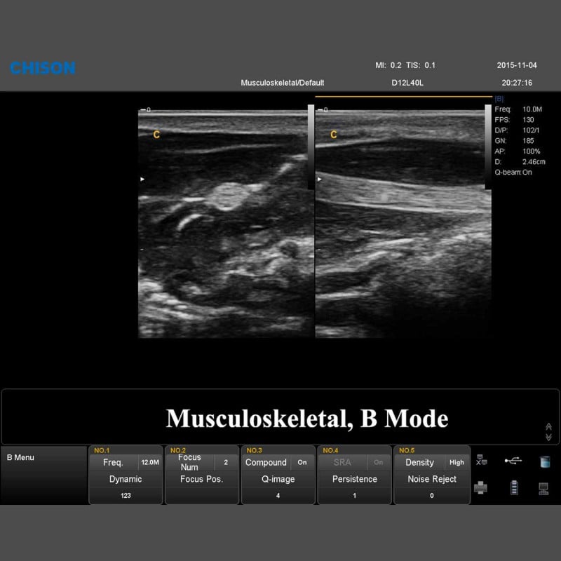
- MSK
- Small Parts
- Urology
- Paediatrics
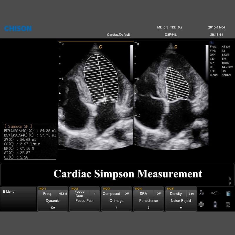
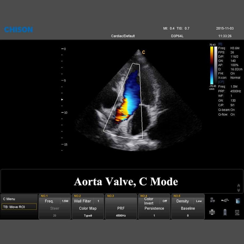
- Cardiology
Technical Specifications
| Dimensions | 806 x 543 x 1360 mm (L x W x H) |
| Weight | 52 kg (without probes) |
| Screen | 21.5" LED colour display; 1920 x 1080 px; brightness & contrast adjustable; multilingual |
| Control Panel | Alphanumeric keyboard, 8 TGC sliders, backlit |
| Probe Connections | 4x |
| Storage Capacity | ≥320 GB integrated hard drive |
| Systems | Image archiving, patient information management, report generation |
| Connections | AC power input: 1; AC power output: 1; On/Off switch: 1; USB port: 6; Ethernet: 1; Remote control: 1; S-Video output: 1; Audio: L, R; DVI: 1; VGA output: 1; Video output: 1; Footswitch connection: 2; Ground connection: 1 |
Overview of Probes
| Convex Probe D3C60L | Linear Probe D7L40L | |
| Application | Abdomen, Obstetrics, Gynaecology, Thorax |
Vascular, Small Organs, Nerves, MSK, Paediatrics, Orthopaedics, Thorax |
| Mean Frequency | 3.5 MHz | 7.5 MHz |
| Number of Elements | 128 | 128 |
| Contact Surface | 73 x 16 mm | 44 x 11 mm |
| Working Modes | B, C, D, M, CPA, DPD | B, C, D, M, CPA, DPD |
| Radius | 60 mm | 40 mm |
| Field of View | 69° | 3.96 cm |
| Scan Depth | 3.2-36.7 cm | 1-14.8 cm |
Share