QBit 9 Ultrasound System
QBit 9 Ultrasound System
Couldn't load pickup availability
- Digital color Doppler ultrasound system
- Advanced technologies (FHI, X-contrast, etc.)
- Professional cardiac package (Stress Echo, CW, PISA and more)
- Includes trolley with four lockable wheels
- Delivery includes convex and linear probe
QBit 9 Ultrasound System
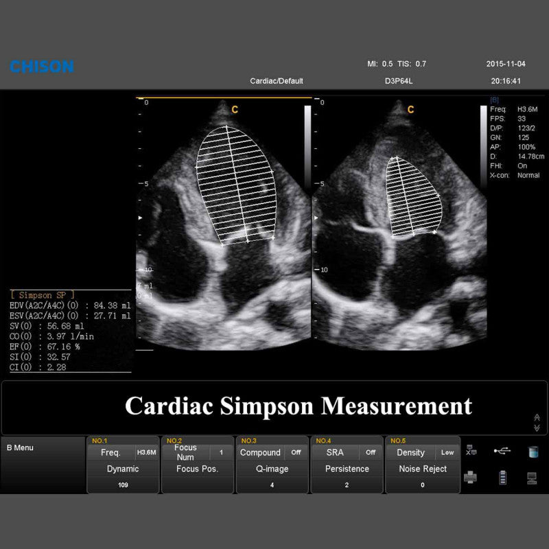
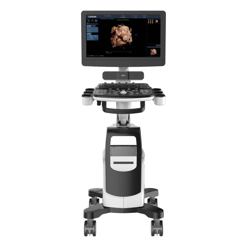
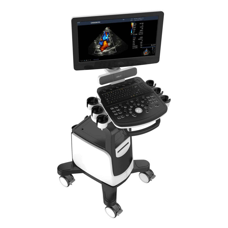
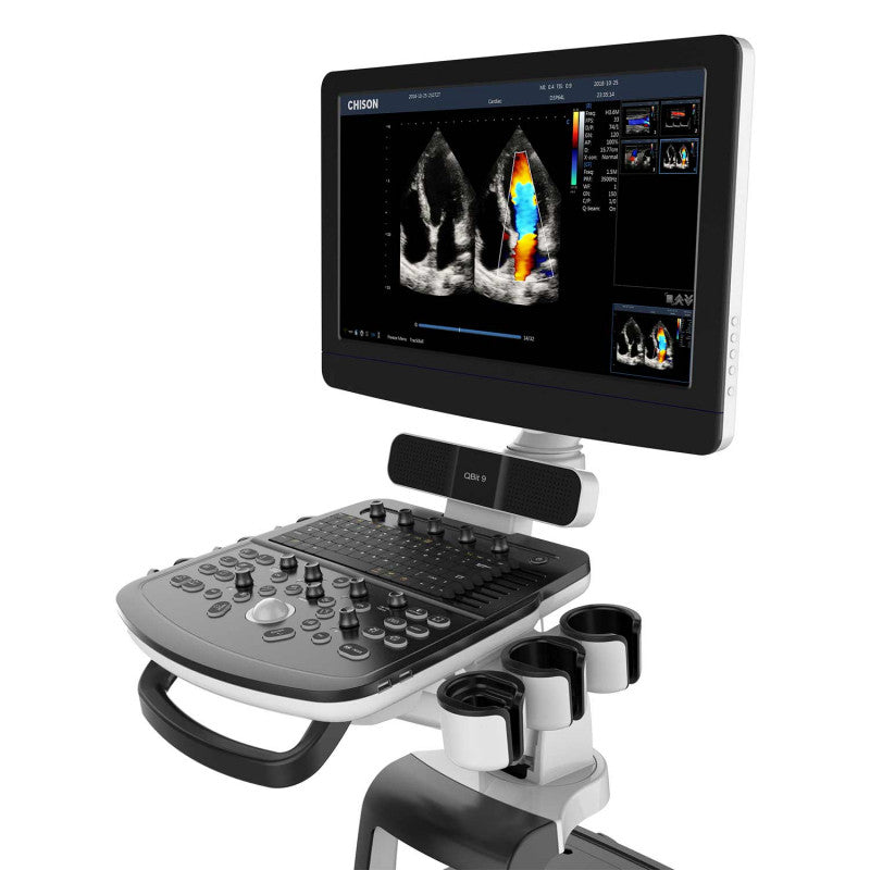
The QBit 9 Ultrasound System from CHISON is an advanced digital color Doppler ultrasound system featuring state-of-the-art technologies such as FHI (Frequency Harmonic Imaging) and X-Contrast. These technologies enable clear and detailed imaging, especially for visualising a variety of tissues. The system provides professional cardiac packages including Stress Echo, CW, PISA and Tissue Doppler Imaging (TDI), specially developed for cardiovascular examinations. Semi-automatic measurements streamline workflows and enhance efficiency.
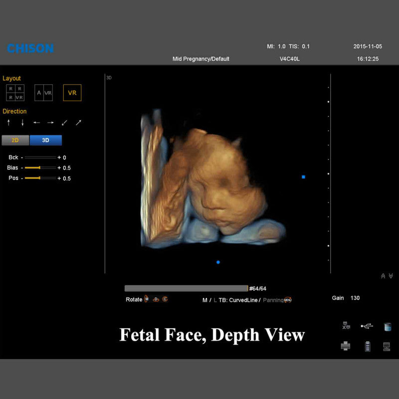
The ultrasound system also includes advanced 4D technologies like Virtual HD and Depth View, enabling even more accurate diagnoses. A standout feature is the 90° swivel 21.5" LED screen, which is up and down foldable, allowing for optimal screen positioning. Various angles for both patient and operator are easily achievable.
The floating keyboard can be rotated from -45° to 45° and is height-adjustable, with backlit keys and TGC buttons with 8 pairs of dials making operation particularly intuitive. A high-quality stereo audio system completes the package. The QBit 9 is also equipped with the innovative Hero Kit for fast and reliable service solutions and includes a built-in dust filter as well as easily accessible paper output. The device trolley with four lockable wheels and the exceptionally low-maintenance design make the system the ideal choice for demanding medical professionals.
Product Details
- QBit 9 Ultrasound System from CHISON
- Digital color Doppler ultrasound system
- Uses advanced technologies (FHI, X-contrast, etc.)
- Includes professional cardiac packages (Stress Echo, CW, PISA, TDI, etc.)
- Semi-automatic measurements
- Offers advanced 4D technologies, such as Virtual HD & Depth View
- 90° swivel 21.5" LED screen
- Screen tilts up and down by 90°
- Optimal screen adjustment to user position
- Various viewing angles for patient and operator
- Intuitively operated control panel and alphanumeric keyboard
- Floating keyboard with left/right rotation from -45° to 45°
- Keyboard with backlit keys and adjustable height
- TGC buttons with 8 dial pairs
- Equipped with a high-quality stereo audio system
- Hero Kit for innovative service solutions
- Built-in dust filter, removable if needed
- Easy access to paper output at the front
- Includes trolley with four lockable wheels
- Exceptionally low-maintenance system
- Extensive probe selection broadens application areas
- Delivered with Convex and Linear probes
- Has four probe slots
- Integrated storage capacity of 500 GB
- Equipped with 4 USB ports
- Optional remote control and footswitch use
- Can be combined with a separately available battery if needed
- VGA and DVI output for external monitors
- LAN output for PC printers, DICOM, and image review stations
- Manufacturer's warranty: 24 months
Advanced Technologies of the QBit 9
| Technology | Features |
|---|---|
| X-contrast | - Adjustable contrast resolution across 3 different levels depending on tissue differentiation. - Regulated via a setting: increase, normal, suppress. |
| FHI | - Utilizes different transmission and reception methods for various patient sizes. - More advanced than traditional THI for better image harmonization. - Improves diagnostic results, especially for obese patients. |
| Q-beam | - Uses Quad-Beam technology instead of Dual-Beam for significantly better images. - Higher image rate ensures better diagnostic reliability and efficiency. |
| Q-flow | - Adaptive color recognition technology. - Automatically differentiates between tissues and adapts the color signal. - Strong improvement in color sensitivity for low-speed flow. |
| SRA | - Algorithm that reduces noise in ultrasound images to improve image quality. - Sharper images with better detail visibility of tissue structures. - Reduces image artifacts and enhances diagnostic reliability. |
| MCI | - Combines ultrasound images from multiple angles for better tissue structure visualization. - Improved resolution and contrast by reducing noise and artifacts. - Clearer, more detailed images that make it easier to identify pathologies. |
| Super Needle | - Enhances needle visibility, e.g., during biopsies or injections. - Improves precision in needle guidance and real-time visualization. |
Advanced Cardiovascular Technologies
PISA:
PISA stands for "Proximal Isovelocity Surface Area", a method for assessing flow convergence to calculate the severity of MR/TR/PR.
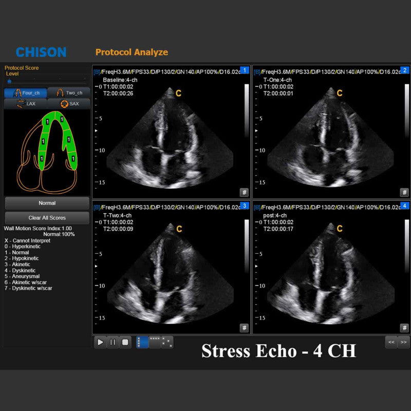
StressEcho:
An echocardiogram is a painless, harmless test that uses high-frequency sound waves to examine the anatomy and function of the heart.
Tissue Doppler Imaging (TDI):
Tissue Doppler Imaging is a novel echocardiographic technique that directly measures myocardial velocity. Systolic TDI measurements assess left- and right-ventricular myocardial contraction function. Diastolic TDI values indicate myocardial relaxation.
Free Steering M Mode:
The cursor line can rotate 360 degrees and be positioned at the desired location; up to three lines can be used for simultaneous measurements.
Applications
Abdomen, Gynecology/Obstetrics, Vascular, Musculoskeletal System (MSK), Small Organs, Urology, Pediatrics, Cardiology
Available Imaging Modes
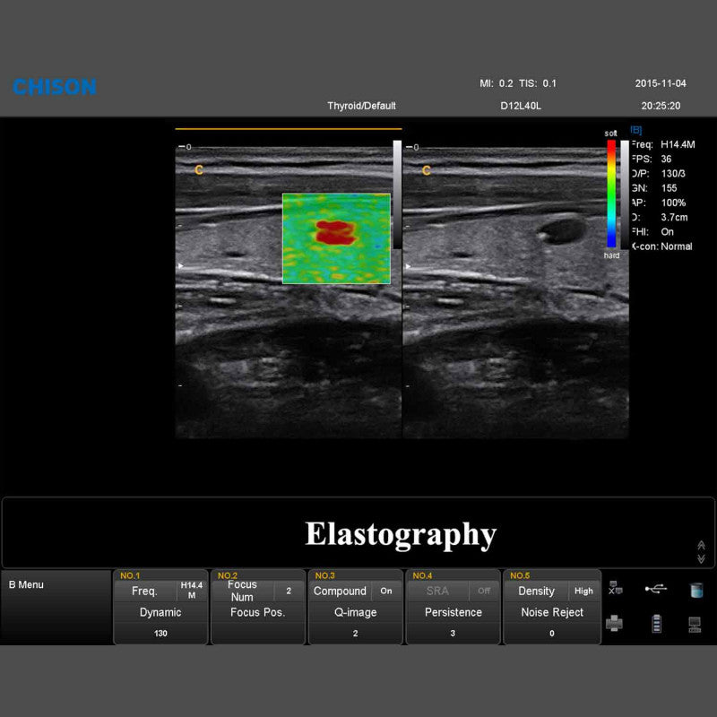
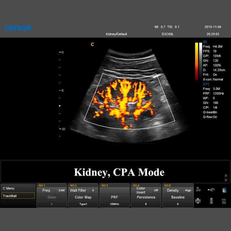
- B, 2B, 4B, B/M, M, CFM, B/BC, PW/CW, PD, Directional PD, Duplex, Instant Triplex, Quadplex, Trapezoidal Imaging, Curved Panoramic Imaging, 2D Control, Chroma B/M/PW/CW, 4D, Virtual HD/Depth View, Control M, Color M, TDI, StressEcho, PISA, Elastography (available with 8 different probes), ECG
Overview of Probes
| Convex Probe D3C60L | Linear Probe D7L40L | |
|---|---|---|
| Application | Abdomen, Obstetrics, Gynecology, Thorax | Vascular, Small Organs, Nerves, MSK, Pediatrics, Orthopedics, Thorax |
| Frequency | 3.5 MHz | 7.5 MHz |
| Number of Elements | 128 | 128 |
| Contact Area | 73 x 16 mm | 44 x 11 mm |
| Radius | 60 mm | 40 mm |
| Field of View | 69° | 3.96 cm |
| Scan Depth | 3.2–36.7 cm | 1–14.8 cm |
Share