Product ID:
SKU:137717
SonoScape S11 Plus ultrasound system without battery
SonoScape S11 Plus ultrasound system without battery
Couldn't load pickup availability
Product overview
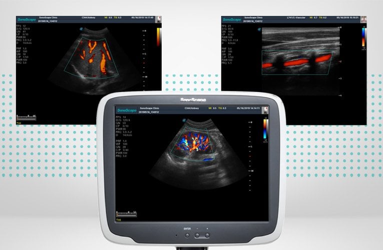
This digital colour Doppler ultrasound system comes with an excellent image quality und is suitable for a broad range of applications in the hospital routine.
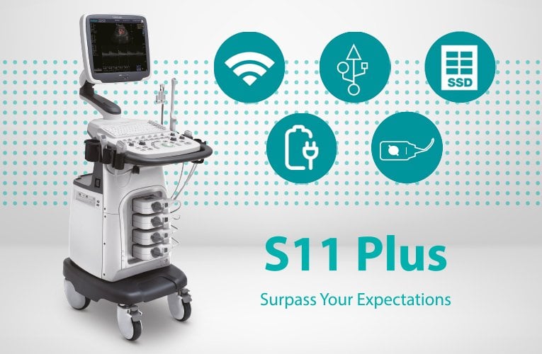
SonoScape S11 Plus
SonoScape S11 Plus is a versatile ultrasound system which is ideal for professional use in various kinds of specialist practices. Delivery includes the linear transducer L741 and the convex transducer C344 which makes it suitable for use in gynaecology and obstetrics. Apart from that, the transducer sockets can be used for examinations of the abdomen, nerves, vessels and small parts.
The SonoScape S11 Plus is alternatively available with a Li-Ion battery. This enables examinations in emergency situations without access to power supplies. The power capacity of the fully charged battery allows for an operating time of over 60 minutes.
Comfortable to use
SonoScape S11 Plus is very comfortable to use thanks to the user-friendly control panel with backlit buttons and multiple defined-keys.
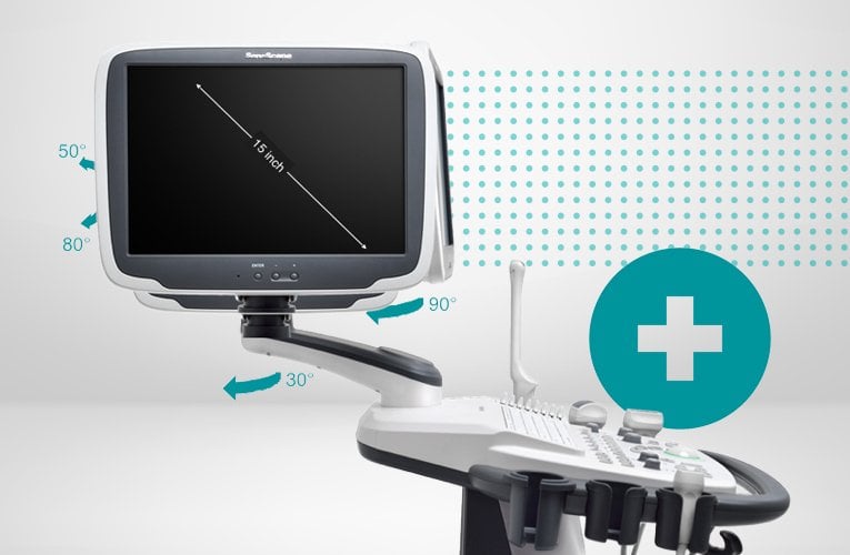
The 15"-LCD colour monitor is equipped with an articulating arm for an ideal view of the examined area. The upper part of the articulating arm is adjustable in height and angle so the monitor can be easily moved to the ideal position. Additionally, the monitor can be tilted to the front, to the back and up to 30° sidewards.
The device comes equipped with 4 transducer ports, 5 transducer holders as well as holders for ultrasonic gel and ultrasonic transducer cable to ensure an optimal workflow. Therefore, there is enough space for the whole equipment and it is easy to switch transducers if needed.
With the help of its four lockable casters and the sturdy handle on the control panel the colour Doppler ultrasound system can be easily adjusted to the desired position.
Optimised image quality
Technical features of the SonoScape S11 Plus for best image quality:
SCI (Spatial Compound Imaging)
With the help of the SCI technology sound waves can be controlled during an ultrasound examination to make them hit the tissue from different, overlapping angles. Thereby, various overlapping scans of an object from different perspectives are registered and then put together in one image. The emerging image shows decreased noise as well as increased boundary and edge detection.
µ-scan technology
µ-scan technology decreases redundant speckle noise and thus ensures high contrast resolution which enhances boundaries and edges and accentuates image features.
PIH (pure inversion harmonic imaging)
PIH technology creates higher resolution combined with a reduction of interfering noise and random noise. This enables clearer structures and enhances image features during ultrasound examinations of small parts and vessels.
Areas of Application
Obstetrics
Gynaecology
Cardiology
Paediatrics
Urology
Abdomen
Foetus
Small parts
Transrectal applications
Transvaginal applications
Peripheral blood vessels
Musculoskeletal system
Musculoskeletal examinations
Product Details
15" LCD colour monitor, resolution: 1024 x 768
Tiltable monitor 30° tot he back, 90° to the front, 30° sidewards
Monitor adjustable in height due to articulating arm (approx. 10 cm)
Brightness and contrast adjustable
User-friendly control panel
Backlit keyboard
Multiple defined-keys
Port options: VGA, USB, network connection, S-video, audio, video, video printer, ECG, mains connection
2 transducers included (linear transducer L741 Line and convex transducer C344)
500 GB HDD hard disk
4 transducer ports
5 holders for ultrasound transducers
1 holder for coupling gel
2 cable holders
4 lockable casters
Alternatively available with integrated battery (enables uninterrupted service of over 60 minutes without mains supply)
Languages available: German, English, French, Italian, Spanish, Russian, Chinese, Norwegian, Portuguese
Foot switch for printing & screen freeze separately available
Dimensions (L x W x H): approx. 52 cm x 130 cm x 72 cm
Weight: approx. 60 kg
Delivery Contents
SonoScape S11 Plus
Linear transducer L741
Convex transducer C344
Power cable
Optional: Li-Ion battery
Display modes
B-mode (2B & 4B
B-mode, panoramic imaging
M-mode
Anatomical M-mode
Colour M-mode
Colour Doppler flow imaging
Power Doppler imaging/directional power Doppler imaging
Pulse wave Doppler imaging
High pulse repetition frequency
Tissue harmonic imaging (THI)
Pulse inversion harmonic imaging
Lateral gain compensation (LGC)
Continuous wave Doppler imaging
Compound imaging
Auto NT / Auto EF / Auto IMT / Auto Trace (PW)
Trapezoid imaging
M-tuning
Freehand 3D
ECG
Tissue Doppler imaging (TDI)
Simultaneous mode (Triplex)
Static 3D/4D imaging
Imaging technologies & software
µ-scan (2D speckle reduction technology)
Multi-beam technology
Vis-Needle (needle visualization enhancement)
Spatial compound imaging (SCI)
Pure inversion harmonic imaging (PIH)
DICOM 3.0
Share